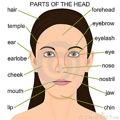
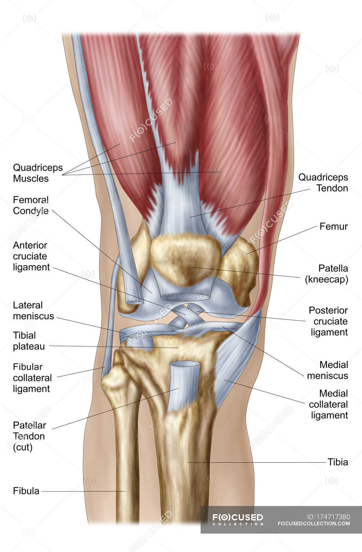
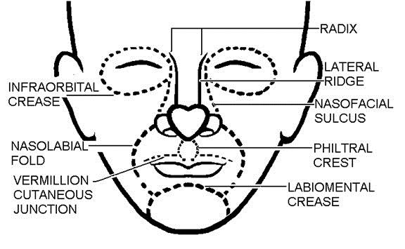
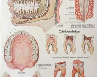
38 face diagram with labels
How to plot a ternary diagram in Excel Adding labels to the tick marks Use the Add Chart Element > Add Labels ( Chart Design tab) to add Data Labels to the A to B axis aligned to the right (Figure 17), then add Data Labels aligned left to the C to A axes. Figure 17: Adding Data Labels to the A-B and B-C axes. By default, Excel will use the Y Value as Data Label. The 14 Facial Bones: Anatomy & Functions - Study.com Here is a labeled diagram of the fourteen facial bones: Facial Bone Names Let's begin with the nasal bones. In this case, we're actually looking at two small bones, which are located just above the...
Ear Anatomy: Understanding the Outer, Middle, and Inner ... Tympanic Membrane or Eardrum. The tympanic membrane, or eardrum is the final hearing organ in the outer ear, separating it from the middle ear. The eardrum collects sound waves and vibrates, passing the sound waves into the middle ear. Most hearing disabilities are caused by trauma or disorders in the tympanic membrane eardrum.

Face diagram with labels
Anatomy of the Epidermis with Pictures - Verywell Health Summary. The epidermis is composed of layers of skin cells called keratinocytes. Your skin has four layers of skin cells in the epidermis and an additional fifth layer in areas of thick skin. The four layers of cells, beginning at the bottom, are the stratum basale, stratum spinosum, stratum granulosum, and stratum corneum. Labelled imaging anatomy cases | Radiology Reference ... This article lists a series of labelled imaging anatomy cases by body region and modality. On this page: Article: Brain. Head and neck. Spine. Chest. Abdomen and pelvis. Parts of a Hammer (With Diagram) - What They are Used for Face The part of a hammer that strikes to different surfaces is called the face. Again, the diameter of a hammer's head varies between different hammers. Some hammers may have a small face whereas some have a large face like the sledge hammer. Claw hammers usually have neither small nor large face, it falls in between. Claw
Face diagram with labels. 12 Cranial Nerves: Functions & Diagram of Locations ... If the facial nerve is damaged, this may impair the ability to make facial expressions on one side of the face. More generally, damage to cranial nerves may result in the following symptoms: Loss of sensation in a part of the face Weakness Numbness of the face Pain Tingling sensation Changes in vision Weak or paralyzed muscles Cat Skeleton Anatomy with Labeled Diagram » AnatomyLearner ... The temporomandibular joint of the cat is a synovial joint and contains an articular disc. Cat skull has a short fascial and palatal region compares to other mammals. The skull is oval elongated in shape, and has strong, highly curved zygomatic bones. There is an incomplete orbital rim in cat skull anatomy. Types of Bones in the Human Body: Skeletal System Labeled ... The 5 main bone types in the human body skeletal system. Labeled diagrams and examples of long bones, short bones, flat bones, sesamoid bones, and irregular bones that make up the foot, hand, skull, cranium, arm, leg, ankle, wrist, hip, and vertebrae or spine. Microscope, Microscope Parts, Labeled Diagram, and Functions The description given below summarize the brief description of microscope parts used to visualize the microscopic specimens such as animal cells, plant cells, microbes, bacteria, viruses, microorganisms etc. The Microscopes parts divided into three different structural parts Head, Base, and Arms. Head/Body: It contain the optical parts in the ...
Circle Diagram: What It Is, Templates & Use Cases - Venngage Create a circular diagram in mere minutes with Venngage's Diagram Maker. 1. Sign up for Venngage with your email, Gmail or Facebook account—it's free! 2. Select one of our professionally designed circular diagram templates or choose a blank canvas. 3. Start editing using our drag-and-drop editor or smart diagram editor. 4. Now's the fun part! Learn the facial muscles with quizzes & labeled diagrams ... Simply fill in the blanks with the face muscles names you remember. DOWNLOAD PDF WORKSHEET (BLANK) DOWNLOAD PDF WORKSHEET (LABELED) How did you get on? If you still need a bit of practice, no worries. This was just a warm-up to get your brain thinking! The next stage is to test yourself using some interactive facial muscles quizzes. Free Printable Parts Of The Body Activity Pack The only thing you need to do to prep this is print out the pack. The first page includes a diagram of a child's body and labels the parts we are exploring with this pack. This has a front side and back side with labels for all of it. The next page shows a girl and a few of the body parts including leg, arm, face, etc. Eye Diagram Quiz - ProProfs Eye Diagram Quiz . 17 Questions | By Bellamiller123 | Last updated: Mar 20, ... Label The Parts Of The Eye. ... Look straight into a mirror, is the length between the side of your face and your eye... Longer than half your eye length. Shorter than half your eye length. Equal to half your eye length.
Facial Bones Anatomy | List & Functions - Video & Lesson ... This image of the skull shows the names of the 14 facial bones. The names of the 14 facial bones are: Inferior nasal concha x 2 Lacrimal bones x 2 Mandible Maxilla x 2 Nasal bones x 2 Palatine... Clavicle - Definition, Location, Anatomy, & Labeled Diagram Clavicle, commonly known as collarbone, is a slender, S-shaped, modified long bone located at the base of the neck. It is the only long bone of the body that lies horizontally. The term clavicle comes from the Latin word ' clavicula ', meaning 'little key', as its shape resembles an old-fashioned key. Also, the bone rotates along its ... Anatomical Position and Directional Terms: Definitions ... Anatomical Directional Terms: Labeled diagram showing superior, defined as above or toward the head. Inferior If we move away from the head, then we are moving inferior. Therefore, inferior is defined as "below or away from the head". You can use the "F" in inferior to think of "Floor", and this can help you remember inferior is toward the floor. Lymphatic System: Functions, Diagram, and Definition Lymph is a fluid connective tissue that flows inside the specialised vessels known as lymphatic vessels. It is a colourless fluid that is a part of the tissue fluid, that in turn, is a part of the blood plasma. The lymph contains very small amounts of nutrients and oxygen but contains abundant carbon dioxide and other metabolic wastes as ...
Head anatomy: Muscles, glands, arteries and nerves - Kenhub Learn and practice the facial muscles more effectively using our facial muscles quizzes and labeled diagrams. There are five main groups of facial muscles, each one consisting of several smaller muscles that are responsible for the movement of a particular region of the face: Orbital group Nasal group Oral group Auricular group Scalp and neck group
Anatomy, Head and Neck, Face - StatPearls - NCBI Bookshelf The most anterior region of the head is the face. The human face is a unique aspect of each individual. The face contains many structures that contribute to the display of emotions, feeding, seeing, smelling, and communicating. One of the most distinguishing qualities of the face is that it is used for personal identity from person to person. Identity is essential since the face is usually the ...
Parts of a microscope with functions and labeled diagram Figure: Diagram of parts of a microscope There are three structural parts of the microscope i.e. head, base, and arm. Head - This is also known as the body. It carries the optical parts in the upper part of the microscope. Base - It acts as microscopes support. It also carries microscopic illuminators.
Structure and Functions of Human Eye with labelled Diagram The External Structure of an Eye. Sclera: It is a white visible portion. It is made up of dense connective tissue and protects the inner parts. Conjunctiva: It lines the sclera and is made up of stratified squamous epithelium. It keeps our eyes moist and clear and provides lubrication by secreting mucus and tears.
Parts of A Check Labeled & Explained (with Diagrams) [2021 ... I'll also show you helpful diagrams with labels so that you can properly identify and understand each part. Table of Contents Parts of the check explained 1. Contact information 2. Date 3. Pay to the order of 4. Transaction amount 5. "Money box" 6. Memo 7. Signature line 8. Routing number 9. Account number 10. Check number Parts of a check FAQ
Muscles of Facial Expression | Anatomy - Geeky Medics The muscles of facial expression (also known as the mimetic muscles) can generally be divided into three main functional categories: orbital, nasal and oral. These muscles are all innervated by the facial nerve (CN VII).¹. These striated muscles broadly originate from the surface of the skull and insert onto facial skin.
Anatomy, Head and Neck, Salivary Glands - StatPearls ... The salivary glands are exocrine glands that make, modify and secrete saliva into the oral cavity. They are divided into two main types: the major salivary glands, which include the parotid, submandibular and sublingual glands, and the minor salivary glands, which line the mucosa of the upper aerodigestive tract and the overwhelming entirety of the mouth [1].
Tennis Court Explained | Diagram Labeled With Dimensions ... In the following diagram, different parts of the tennis court are labeled with dimensions. Diagram of Tennis Court. In this diagram: Tennis Court Explained with Dimensions and Labels ... The ad side is on the left side when you face the net. The player keeps switching from deuce to add until the game ends. 8. Center Mark.
Horse Skeleton Anatomy - Osteological Features of Bones ... Horse skeleton anatomy diagram Few special osteological features from the axial and appendicular skeleton of a horse - The skull of a horse is long and four-sided. You will find an extensive foramen lacerum in the horse skull. There is no cornual process in horse skull. The fusion between the two haves of the mandible is complete.
Illustrations and diagrams of the 12 pairs of cranial ... This human anatomy module is about the cranial nerves. It consists of 15 vector anatomical drawings with 280 anatomical structures labeled. It is intended for the use of medical students working on human anatomy, student nurses, physiotherapists, electro-radiological technicians and residents - especially those working in neurology, neurosurgery, otolaryngology - and for any physician ...
Anatomy of the face and neck (MRI) - e-Anatomy - IMAIOS The bones of the face and neck were labeled using different colors to facilitate comprehension. The bone structures are rather more difficult to view on a weighted MRI T2 than on a CT-Scan: for more details on the bones of the face, please refer to the e-Anatomy module "Face-CT-Scan". The teeth were numbered using the FDI World Dental ...
Parts of a Hammer (With Diagram) - What They are Used for Face The part of a hammer that strikes to different surfaces is called the face. Again, the diameter of a hammer's head varies between different hammers. Some hammers may have a small face whereas some have a large face like the sledge hammer. Claw hammers usually have neither small nor large face, it falls in between. Claw
Labelled imaging anatomy cases | Radiology Reference ... This article lists a series of labelled imaging anatomy cases by body region and modality. On this page: Article: Brain. Head and neck. Spine. Chest. Abdomen and pelvis.
Anatomy of the Epidermis with Pictures - Verywell Health Summary. The epidermis is composed of layers of skin cells called keratinocytes. Your skin has four layers of skin cells in the epidermis and an additional fifth layer in areas of thick skin. The four layers of cells, beginning at the bottom, are the stratum basale, stratum spinosum, stratum granulosum, and stratum corneum.


Post a Comment for "38 face diagram with labels"